For more information call
The Wellington Hospital: 020 7483 5589
Fortius Clinic: 0845 853 1000 The Yorkshire Clinic: 01274 621600 |
|
Hip Anatomy
The Hip Joint
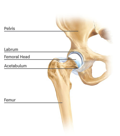
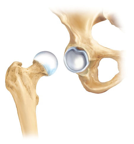
The hip is a ball and socket joint. The ball (head of the femur) is kept in the socket (acetabulum, part of the pelvis). The socket consists of a bony part, which is surrounded by a thick soft tissue rim known as the labrum. There are multiple ligaments which form part of the envelope of soft tissue around the hip, and which help keep the ball in the socket and stabilise the joint.
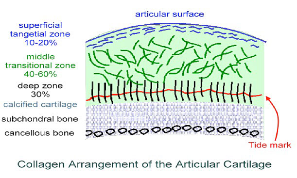
The hip joint, like most joints, relies on a surface coating of frictionless articular cartilage to enable smooth pain-free movement .
Articular cartilage is very smooth and it can withstand high compression forces for its weightbearing function. Articular cartilage derives its powers from a very organized and efficiently constructed microanatomy. At the base, horizontal fibers are firmly adherent to the subchondral bone. The layers above contain macromolecules which bind water. This hydration gives the cartilage its typical springiness needed for shock absorbency. The top layer has tangentially orientated collagen fibers which serve as a skin to protect the cartilage from shearing forces.
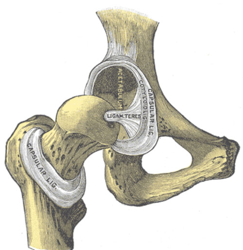
The labrum is a soft tissue fibrocartilage ring around the acetabulum. It is horseshoe shaped and the ends are connected via the transverse ligament. The labrum increases the depth of the socket. It has an important biomechanical and biological function. The labrum seals off the joint keeping the fluid inside and also increases the stability by creating a negative pressure within the joint. The suction seals aid with the nutrition of the articular cartilage therefore decreasing the chondral degeneration. With a deficient labrum the suction seal on the femoral head decreases and outwards movement of the femoral head can occur. Gradual detachment of the labrum and the wearing down of the articular cartilage can occur with femoroacetabular impingement. ( labral tear - labral repair - surgical result )
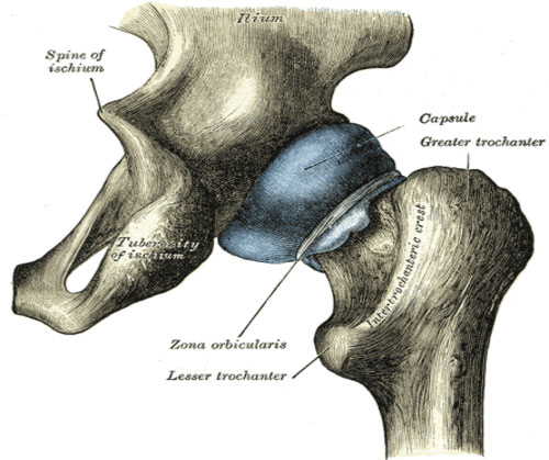
Hip capsule and ligaments
There are multiple ligaments that form part of the envelope of soft tissue around the hip, and help keep the ball in the socket and stabilise the joint. The ligaments are reinforcements of the hip capsule. The most important one is the Ilio femoral ligament at the front of the hip joint, which is also the strongest ligament in the human body.
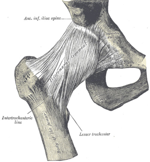
The function of the hip ligaments is to stabilise the hip joint, but also to control movement and restrict the joint from going into extreme positions. The iliofemoral ligament is Y shaped and resists external rotation (prevents the foot from falling out) and internal rotation. In some sports that require a lot of rotation, the iliofemoral ligament can be stretched out creating symptoms of hip instability.
Another ligament that reinforces the hip capsule, is the zona orbicularis. This consists of circular fibers, which form a sling around the femoral neck. This structure is important to resist distraction and axial forces to the hip joint.
The ligamentum teres is a round ligament that connects the femoral head with the acetabulum. The ligament contains a blood vessel, which contributes to the blood supply of the femoral head during childhood. The role of this ligament is less significant after childhood.